Created by - Rigomo Team
The general approach to the trauma patient
In the first forty years of life, trauma is the main cause of death. Each year, it causes 10 million disabilities and 60 to 80 million injuries. Survival for trauma patients depends on prompt diagnosis and treatment. Traumatic injuries such as subdural and epidural hematomas, hemopneumothorax, tension pneumothorax, pericardial tamponade, burst spleen, lacerated liver, pelvic fractures, and aortic disruption can result in death within minutes.PRIMARY SURVEY: The primary survey consists of the “ ABCs”.1. A—airway and cervical spine control: The patency of the airway is assessed, with the cervical spine immobilized. The presence of a cervical spine injury should be assumed until proven otherwise.2. B—breathing and ventilation: The chest is auscultated and inspected, and the quality, depth, and rate of respirations are noted.3. C—circulation and hemorrhage control: Capillary refill, pulses, and the color of the skin should be assessed. Active hemorrhages should be identified and treated with direct pressure or surgical repair. Two large-bore (14- to 16-gauge) intravenous catheters are inserted and intravenous fluids (lactated Ringer or normal saline) are started. Set up a pulse oximeter and a heart monitor for the patient.4. D—disability: A rapid neurologic assessment is performed, assessing the pupils for size, equality, and reactivity, and a Glasgow coma scale.5. E—exposure: The patient should be completely undressed then warm blankets applied to prevent hypothermia.PATIENT HISTORY: Whenever possible, an AMPLE history (allergies, medications, past medical history, last meal, and events surrounding the injury and its mechanism) should be obtained from the patient and pre-hospital personnel. X-rays (chest and pelvis) and lab tests are now ordered (complete blood count, basic metabolic panel, pregnancy test, type and screen, and prothrombin time/international normalized ratio).SECONDARY SURVEY: This is a head-to-toe complete physical exam, examining each region systematically. During the primary or secondary survey, focused assessment sonography in trauma (FAST) exam can be done in 2 minutes to risk stratify the trauma patient.
More detailsPublished - Thu, 08 Sep 2022

Created by - Rigomo Team
Stages of Wound Healing
1. Immediate response to tissue injury— Vasoconstriction and tissue contraction occur.— Platelets aggregate and a clotting cascade is activated.— Once hemostasis is complete, the release of vasoactive amines causes capillary dilation, and an exudate forms.2. Inflammatory phase — Granulocytes and lymphocytes accumulate to control the growth of bacteria and prevent infection.— Immunoglobulins contribute to the control of infection.— Macrophages phagocytize debris, encourage collagen deposition, and stimulate neovascularization.3. Epithelialization— Within 12 hours of injury, cells of the stratum germinative are activated, and within 24 hours, initial epithelialization may be complete.4. Neovascularization— New blood vessels form and bring nutrients and oxygen to the healing wound.— Neovascularization begins on day 3 and peaks on day 7.— By day 21, the process is complete and the new blood vessels withdraw as the tissue matures.5. Collagen synthesis— Fibrocytes initially lay down a disorganized pattern of collagen.— Over months to years, the matrix is remodeled to form an organized meshwork of collagen.6. Wound contraction— Myofibroblasts are responsible for wound contraction, in which the initial scar contracts to a smaller size.
More detailsPublished - Fri, 09 Sep 2022

Created by - Rigomo Team
What is Acute Respiratory failure (ARF)?
Acute Respiratory failure (ARF) is a syndrome, characterized by the inability of the system to support effective and continuous gas exchange and elimination of carbon dioxide in the atmosphere. ARF is a medical emergencyThe syndrome is recognized to have contributed to increased mortality globally, and a high rate of ICU admission. Most typical causes of ARF embrace acute respiratory illness, chronic impending respiratory organ failure, chronic coronary insufficiency, and acute respiratory distress syndrome. There are 2 main types of ARF: Hypoxemic respiratory failure: Lack of oxygen in the blood, but levels of carbon dioxide are almost normal.Hypercapnic respiratory failure: Presence of too much carbon dioxide in the blood, and near normal or lack of oxygen in the blood.Symptoms of ARF.o Difficulty breathingo Cyanosis [bluish coloration in the skin, fingertips, or lips]o Rapid breathingo Restlessness, confusiono Drowsinesso Irregular heartbeatso Excessive sweating Diagnosis and TreatmentIn ARF emergency measures of diagnosing and treatment ought to be enforced at the same time to forestall the progression and complications of respiratory The diagnosis of ARF is usually suspected based on the symptomatic profile of the patient, the body’s oxygen and carbon dioxide levels level, an arterial blood gas test, and a chest X-ray/CT Scan to find out the lung abnormality. General principles of treatment of ARF patients are similar regardless of the underlying metabolism pathology. These include: 1) Check for patency of Airways, and look for breathing and circulation2) Oxygen supplementation through the mask or nasal tubing to maintain adequate oxygen levels. 3) Tracheostomy is needed if a patient is on prolonged ventilator supportImprovements in current medical technologies, better respiratory support after early detection, and immediate intervention have contributed to a decrease in the overall morbidity and mortality from ARF.
More detailsPublished - Fri, 09 Sep 2022

Created by - Rigomo Team
Pulmonary Embolism – Diagnosis & Treatment
Pulmonary embolism is usually tough to diagnose, particularly in folks who have an underlying heart or respiratory organ illness. For that reason, your doctor can probably discuss your symptoms, do a physical examination, and order one or more tests for confirming the diagnosis.Blood tests that may include o The D-dimer [clot-dissolving substance] test o High levels of D-dimer indicate an exaggerated chance of blood clots, though several other factors may also cause high D-Dimero ABG analysis to know the status of oxygen and carbon dioxide [CO2] in your blood. A clot can decrease the oxygen levels in your blood.o To assess the clotting disorder [if any] Chest X-rayThis noninvasive check shows pictures of your heart and lungs on film. Though X-rays cannot diagnose embolism, however, they will help rule out other conditions that mimic the illness. UltrasoundA noninvasive check called duplex imaging (sometimes referred to as duplex scan or compression ultrasonography) uses sound waves to scan the veins in your thigh, knee, and calf, and typically in your arms, to visualize for deep vein blood clots.The absence of clots reduces the chance of deep vein occlusion. If clots are present then the treatment is started right away.CT pulmonary angiographyCT pulmonary angiography creates 3D pictures that may discover abnormalities like embolism inside the arteries in your lungs. In some cases, a contrast medium is given intravenously so to stipulate the respiratory organ arteries while scanning.Ventilation-perfusion scan (V/Q scan)When there's a requirement to avoid radiation exposure or distinction from a CT scan because of a medical condition, a V/Q scan could also be performed. During this check, a tracer is injected into a vein in your arm. The tracer maps blood flow (perfusion) and compares it with the flow of air to your lungs (ventilation) and may confirm whether or not blood clots are inflicting symptoms.Pulmonary AngiogramThis check provides a transparent image of the blood flow within the arteries of your lungs. it is the most correct way to diagnose embolism, however, it needs a high degree of precision to administer and has probably serious risks, it's always performed once alternative tests fail to produce a definitive diagnosis.MRIMRI may be a medical imaging technique that uses a force field and computer-generated radio waves to form elaborate pictures of the organs and tissues in your body. This imaging is typically reserved for pregnant ladies (to avoid radiation to the fetus) and people whose kidneys are damaged by dyes utilized in other tests. TreatmentTreatment of embolism is aimed toward keeping the blood clot from getting larger and preventing new clots from forming. Prompt treatment is crucial to stop serious complications or death.MedicationsMedications embrace different kinds of blood thinners and clot dissolvers.o Blood thinners (anticoagulants). These medications stop existing clots from enlarging and new clots from forming whereas your body works to interrupt the clots. Heparin is the most common anticoagulant that can be given through the vein or injected under the skin.o Clot dissolvers (thrombolytics). Whereas clots sometimes dissolve on their own, typically thrombolytics given through the vein will dissolve clots quickly. The clot-busting medications can cause severe bleeding, they are reserved for life-threatening conditions.Surgical and alternative procedureso Clot removal. If you have got a massive, dangerous clot in your lungs, your doctor might consider removing it via a skinny, versatile tube (catheter) through your blood vessels.o Vein filter. A filter is positioned in inferior vena cava, a vein that leads from your legs to the right side of your heart. This filter will facilitate keep clots from going to your lungs. This procedure is often reserved for folks that cannot take anticoagulant medication or have had continual clots formation despite the use of anticoagulants. Some filters will be removed when they are no longer needed.Ongoing care Because you'll be in danger of another deep vein occlusion or embolism, it is very important to continue treatment, like remaining on blood thinners and be monitored frequently as advised by your doctor.
More detailsPublished - Sat, 10 Sep 2022

Created by - Rigomo Team
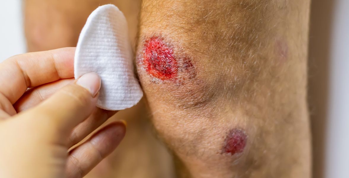
How to Evaluate Wounds in the Emergency Department
The goals of wound care in the emergency department (ED) are to prevent infection, restore function, and restore physical integrity.EXAMINATION OF THE WOUND allows the physician to assess the level of care required. Attention to the area of injury, and its underlying structures, guides the physician in deciding whether the wound can be treated in the ED or requires a surgeon.HISTORY OF PRESENT INJURY1. Contamination of the wound: Wounds with high concentrations of bacterial contamination (e.g., by feces, saliva, or organic matter) need extensive debridement and irrigation. Some of these wounds may have such extensive contamination that they will require delayed closure.2. Age of injury: The golden period of wound care is generally considered to be less than 6 to 8 hours following injury. Between 8 and 12 hours, some wounds can be closed without a significant additional risk of infection. It is best to not close wound that is greater than 16 hours unless there is a cosmetic issue. In contrast, in a heavily contaminated wound of the foot, closure may not be safe in as little as 3 hours postinjury.3. The extent of injury: Inspect all wounds for injuries to deep structures like tendons, nerves, and blood vessels. Pull wounds open and debride so the entire wound can be seen.4. Past medical historya) Past medical history: Diabetes, immunosuppression (e.g., caused by steroids, AIDS, cancer treatment), and alcoholism are examples of conditions that affect wound healing. These conditions lead to slower healing and higher infection rates. In the presence of such conditions, sutures should be left in longer and prophylactic antibiotics should be considered.b) Age of the patient: Patients younger than 2 years and older than 50 years have higher rates of infection.c) Smoking status: Tobacco use decreases peripheral blood flow and increases the risk of other vascular injuries.d) Nutritional status: Patients with severe nutritional deprivation have slower wound healing and higher infection rates. Supplemental nutrition may be necessary for these individuals.e) Medications: Steroids and immunosuppressive medications may slow healing and increase infection rates. Aspirin, antiplatelet agents, and warfarin may cause an accumulation of blood in wounds that are primarily closed, causing swelling and possible infection.f) Tetanus immunization: Tetanus is a highly preventable and potentially fatal disease. All patients with wounds should be questioned as to their immunization status.GENERAL INSPECTION1. Description of wounds: Document length, depth, ability to visualize the base, and shape (stellate, linear, jagged, flap). The entire wound should be visualized. If it is covered by matted blood or hair, gently clean if off.2. Functionality of wounded area: The initial inspection is used to evaluate functioning of the area as well as distal functioning. Attention to neurologic function (i.e., sensation, reflexes, strength) prior to the use of local anesthesia is important. Muscle movement and tendon function should be evaluated through the entire range of motion. Vascular integrity should be evaluated (i.e., temperature, color, capillary refill, pulses).
More detailsPublished - Sat, 10 Sep 2022

Created by - Rigomo Team
ASTHMA – DIAGNOSIS & TREATMENT
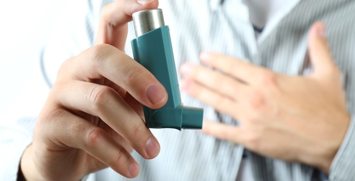
DiagnosisFor adults and youngsters who have recently been diagnosed with Asthma[five years], PULMONARY FUNCTION TESTS are routinely checked to know how well the lungs are operating. Poor results on the tests indicate that respiratory disease is not well controlled. These tests also tell how well the treatment plan is workingLUNG FUNCTION TESTS include:o Peak flow.Your doctor might take a peak flow reading after you are available for a scheduled visit or emergency treatment throughout the treatment. This check measures however quickly you'll exhale. You’ll additionally use a peak flow meter to observe how well your respiratory organ can operate.o The results of this check are called peak breath flow (PEF). A peak flow check is finished by processing into a mouthpiece as quickly as you can with one breath (expiration).o Spirometry. Throughout spirometry, you are taking deep breaths and forcefully exhale into a hose connected to a machine referred to as a SPIROMETER. o It measures§ Amount of air one can breathe in and out. § How fast one can empty the air out of the lungs.o Nitric oxide activity. o This measures the amount of nitric oxide gas out of the body through exhalation as nitric oxide levels sometimes increase in patients with asthma. High gas readings indicate inflammation of the cartilaginous respiratory tubes.o Pulse oximetry. o This check measures the oxygen levels in your blood. It's measured through the tip of the finger TreatmentIf your doctor has diagnosed asthma, always follow his instructions and follow the action plan as asthma can not be cured but can be managed effectively by medicationsIt constituteso Long-term asthma control medications to keep asthma under controlo Inhaled corticosteroidso Leukotriene modifiers.o Combination inhalerso Theophyllineo Quick-relief (rescue) medicationso Short-acting beta-agonists.o Anticholinergic agentso Oral and intravenous corticosteroids.A quick-relief inhaler can help ease the symptoms quckly in case of an asthma flare-up
More detailsPublished - Mon, 12 Sep 2022

Created by - Rigomo Team
Platelet Abnormalities
Platelet abnormalities can be categorized as either disorder of dysfunction (for which transfusion therapy may be futile) or disorders of quantity (for which transfusion is beneficial).1. Decreased platelet production may be seen in patients with marrow dysfunction caused by infiltration, infection, drugs, or radiation.2. Increased platelet destruction is seen in patients with idiopathic thrombocytopenic purpura or TTP, hemolytic–uremic syndrome, DIC, or viral infections.3. Splenic sequestration of platelets can occur in hypothermic patients or those with hypersplenism (e.g., with portal hypertension).4. Platelet loss can be seen with hemorrhage or hemodialysis.5. Thrombocytosis, seen in patients with polycythemia vera, splenectomy, or malignancy, may result in coagulopathy when platelet counts exceed 1 million/mm3.CLINICAL FINDINGS1. Asymptomatic: Incidental thrombocytopenia may be found by laboratory testing in asymptomatic patients.2. Hemorrhage: Patients with significant platelet dysfunction may have some form of mucocutaneous hemorrhage, and extreme platelet disease may result in catastrophic cerebrovascular hemorrhage.— The presence of petechiae, ecchymosis, or purpura may provide clues to the presence of thrombocytopenia.— Mucocutaneous bleeding is suspicious for platelet abnormality.3. Splenomegaly: The finding of splenomegaly on abdominal examination may help define the cause.EVALUATION: Laboratory assessment includes a hematocrit, platelet count, and coagulation profile. Measurement of the bleeding time to assess platelet function is usually performed in a specialized laboratory. The bleeding time usually becomes abnormal with platelet counts lower than 50,000/mm3 (normal counts range from 150,000 to 450,000/mm3).THERAPY1. Platelet administration is indicated for all patients with platelet levels below 10,000/mm3 and many patients with platelet levels below 50,000/mm3. Administration of platelets may be counterproductive in some patients with the hematologic disease (e.g., TTP). Consultation should be obtained before transfusing platelets in such patients.2. Other therapies are necessary when thrombocytopenia is caused by platelet destruction. Prednisone is often indicated for patients with TTP, idiopathic thrombocytopenic purpura, or hemolytic–uremic syndrome.
More detailsPublished - Tue, 13 Sep 2022

Created by - Rigomo Team
How do I know if my kidney disease is getting worse?
Some symptoms are gradual, whereas others come quickly, therefore it’s vital to get the tests done and always stay in touch with your doctor to manage your kidney disease, even though you are feeling fine. Here’s what to look out for: o Lower Back Pain Or Flank Paino Fatigue Or Weaknesso Swelling, Particularly In Your Hands Or Feet, Or Beneath Your Eyeso A Bad Taste in Moutho Feeling Coldo Poor Concentrationo Shortness Of Breatho Itching Skino Cramping In Hands And Legso Nausea /vomiting These symptoms are proof that you might be developing secondary conditions as a result of your kidney damage. These conditions will include: o Anemiao Anemia is a low amount of hemoglobin or lack of healthy red blood cells that are responsible for carrying oxygen from the lungs to the body. o Symptoms of anemia embody fatigue, dizziness, and pale skin. o Mineral and bone disordero Bone mineral density is reduced due to an imbalance in calcium and phosphate o Symptoms might embody pain in your bones and joints o Fluid overloado Fluid overload happens once your kidneys can not maintain the correct quantity of fluids and there's fluid accumulation in your body.o Symptoms might embody swelling, particularly in your feet and legs, weight gain, and shortness of breath. o Metabolic acidosiso Metabolic acidosis happens when acid builds up in your body because your kidneys aren't functioning properly.o Symptoms might embody headaches, dizziness, nausea, and loss of appetite Be sure to talk to your doctor concerning any new or worsening symptoms, or if you believe you're developing a secondary condition as a result of a worsening disease Symptoms of worsening kidney disease are different for every person. Your laboratory test results, together with eGFR and simple protein values, will facilitate establishing your stage of kidney damage and tracking its progression.
More detailsPublished - Tue, 13 Sep 2022

Created by - Rigomo Team
Yoga for kidney health: how this works
It is very essential to take care of all organs of the body. Kidneys remain the essential organ to be taken care of.Kidneys perform the very important perform of purifying and detoxifying the blood by filtering the waste from it along with maintaining fluid balance, and secretion of hormones Kidneys are also responsible for maintaining acid-base equilibrium or maintaining the amount of acid and bases within the body. Kidneys are one of the foremost important organs within the body and must be cared for. A World Health Organization (WHO) analysis has found that just about 1,000,000 lives are lost worldwide, every year due to kidney failure. Moreover, high blood pressure levels and diabetes are considered the risk factors for kidney damage. Yoga for kidney healthYoga poses stimulate various organs in such a way that their functions are improved. Commence with these straightforward yoga poses to attain the health of your kidneys. These poses affect different components of the body similarly. Thus, if you follow yoga for kidney health, you're serving the complete body. The yoga asana is performed to reach a state of extended relaxation. They conjointly assist in managing fluid balance within the body. This allows sound sleep schedules and thence imparts rest. A well-rested body is healthier to perform functions like the regulation of water within the body.Yoga keeps the internal organs healthy, balances the assorted systems within the body, and effectively controls the strain on the body and mind. Several yoga asanas and pranayamas facilitate internal cleansing rituals that make sure the flushing of toxins from the body. This conjointly ensures normal blood circulation throughout the body, keeping it active and energetic. Here are some healthy yoga asanas mentioned for improving kidney health o Ardha Matsyendrasana (Sitting 0.5 Spinal Twist) - The sitting 0.5 spinal twist stimulates the kidneys and liver. It conjointly improves the body’s immunity level.o Salamba Bhujangasana (Sphinx Pose)The sphinx creates stretches and strengthens the abdominal organs. It conjointly raises the immunity level of the body.o Bhujangasana (Cobra Pose)The pose stimulates the abdominal organs and relieves the body of stress and fatigue. It also improves immunity.o Paschimottanasana (Two-Legged Forward Bend)The two-legged forward bend stimulates the kidney, improves digestion, and relieves discomfort.o Setu Bandhasana (Bridge Pose)The bridge pose stimulates the abdominal organs. It helps maintain pressure levels and relieves the body of stress. o Naukasana (Boat Pose) The boat creates strengthens and stimulates the abdominal organs. It conjointly helps improve digestion and alleviates stress
More detailsPublished - Fri, 16 Sep 2022
Search
Popular categories
Health and Wellness
231Skill Development
7Technology
5Community Impact
2Success story
2Creativity
1Latest blogs

DeepSchool: The Story of an Idea That Refused to Sit Still
Tue, 02 Dec 2025

Transforming Emergency Care: The Story Behind Rigomo's Revolutionary PPMMP Course
Sun, 12 May 2024

Empowering Rural Healthcare: How Pogiko's AI is Bridging the Gap in Medical Services
Thu, 25 Apr 2024
Write a public review